Live cell imaging a laboratory manual download
Our innovative instruments open new perspectives into live cell imaging and cell kinetic analyzis. IPRASENSE’s label-free time-lapse imaging technology offers a versatile solution for monitoring cell culture inside your incubator. The unmatched extra large field of view and the insensitivity to focus provide a robust real-time analysis of
imaging a laboratory manual Dec 11, 2019 Posted By Danielle Steel Media Publishing TEXT ID e27c59cf Online PDF Ebook Epub Library laboratory manuals basic methods in microscopy protocols and concepts from cells a laboratory manual drosophila neurobiology a laboratory manual live cell imaging a
MRI–CIA MRI Cell Image Analyzer, developed by the Montpellier RIO Imaging facility (CNRS), is a rapid image analysis application development framework, adding …
Download 184KB. Mapping brain cell types with CARTANA in situ sequencing on the Nikon Ti2-E microscope. Download 6.21MB. A1R HD25: the latest in resonant scanning technology allows new live-cell imaging approaches. Download 1.37MB. Increasing Data Collection and Fidelity by Maximizing Confocal Field of View. Download 335KB. See All
Image live cells or chase with spectrally distinct ligand prior to imaging. Figure 10.2. Overview of protocol for live-cell and fixed-cell imaging using the HaloTag®Technology.This technology allows labeling and analysis of HaloTag®fusion proteins by live- or fixed-cell imaging, gel-based fluoroimaging, Western blotting and capture of HaloTag®
Nanolive develops a high speed, high resolution and label-free live cell imaging microscope to look instantly inside living cells in 3D!
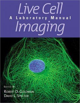
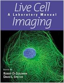
Goldman, R. D., Spector, D. L. (2005) Live cell imaging : a laboratory manual. Cold Spring Harbor Laboratory Press, Cold Spring Harbor, New York. ISBN 0879696834
Live-cell imaging can also involve time-lapse methods, which provide a biologist with dynamic information by periodically taking images over time, showing, for example, how a protein moves in a membrane over time. In fact, cellular dynamics provides a keen area of interest. Applying live-cell imaging on a cellular level exposes biomolecular
Goldman, R. D., Swedlow, J. R., Spector, D. L. (2010) Live Cell Imaging: A Laboratory Manual. Cold Spring Harbor Laboratory Press. ISBN 9780879698935
imaging a laboratory manual Dec 10, 2019 Posted By Jir? Akagawa Media Publishing TEXT ID e27c59cf Online PDF Ebook Epub Library laboratory manuals basic methods in microscopy protocols and concepts from cells a laboratory manual drosophila neurobiology a laboratory manual live cell imaging a
Download Citation Live Cell Imaging, A Laboratory Manual, 2nd Edition. Robert D. Goldman, Jason R. Swedlow, and David L. Spector (Eds.) Live Cell Imaging, A
Access BioTek’s Customer Resource Center to search frequently asked questions and discussion groups for imaging & microscopy, detection, liquid handling, robotics and software. If you’re already a customer, login for full access to participate in peer-to-peer discussions, download user manuals and software, and view order or product information specific to you!
The second edition of Live Cell Imaging: A Laboratory Manual expands upon and extends the collection of established and evolving methods for studying dynamic changes in living cells and organisms presented in the well-known first edition.
Live Cell Imaging A Laboratory Manual
Live cell imaging a laboratory manual – ECU Libraries
Extract meaningful data that leads to new insights and enables faster and more confident decision making. Our powerful image analysis software makes it easy for you to generate and analyze image data from cellular samples and in vivo models so you confidently move your research forward.. High Content Screening: Turn your cellular data into knowledge with our high content analysis software
IncuCyte® Live-Cell Analysis Systems are real-time quantitative live-cell imaging and analysis platforms that enable visualization and quantification of cell behavior over time, by automatically gathering and analyzing images around the clock within a standard laboratory incubator. This enables researchers to make time-lapsed, kinetic
Today cellomics is recognized as a discipline of quantitative cell analysis that is also known as high-content screening (HCS) or high-content analysis (HCA).HCA comprises a powerful combination of fluorescence microscopy, image processing, automated cellular measurements and informatics tools that has enabled fundamental discoveries in biological research.
Mar. 19, 2018 Press release “High-speed super resolution for live cell imaging N-SIM S Super Resolution Microscope” Feb. 28, 2018 Press release “Nikon enters strategic business cooperation for cell-related fields with Berkeley Lights Inc of the United States”
Key words: CLSM, Multi-photon, TIRF, Live-cell imaging, Spinning disk Summary Live-cell microscopy – tips and tools Melanie M. Frigault1, Judith Lacoste2,3, Jody L. Swift4 and Claire M. Brown2,* 1Molecular Oncology Group, McGill University Cancer Centre, Montreal, Canada 2McGill University Life Sciences Complex Imaging Facility, Department of
Not Available adshelp[at]cfa.harvard.edu The ADS is operated by the Smithsonian Astrophysical Observatory under NASA Cooperative Agreement NNX16AC86A
Bimolecular Fluorescence Complementation (BiFC) Analysis of Protein Interactions in Live Cells. Tom K. Kerppola; Adapted from Imaging: A Laboratory Manual (ed. Yuste). CSHL Press, Cold Spring Harbor, NY, USA, 2010. Abstract. Bimolecular fluorescence complementation (BiFC) analysis enables direct visualization of protein interactions and modifications in living cells. It is based on the
Read and Download PDF Ebook life science grade 12 paper 1 november 2010 at Online Ebook Library. Get life science grade 12 paper 1 november 2010 PDF file for free from our online library PDF File: life science grade 12 paper 1 november 2010. LIFE SCIENCE GRADE 12 PAPER 1 NOVEMBER 2010 PDF [PDF] LIVE CELL IMAGING A LABORATORY MANUAL
Live Cell Imaging: A Laboratory Manual. Cold Spring Harbor Press, Cold Spring Harbor, New York, 631 pages (2005). Featuring over 30 chapters written by noted experts, this reference manual is perhaps the most important single source for information related to a wide spectrum of topics in live-cell imaging. The chapters address advanced
Live Cell Imaging: A Laboratory Manual D. Spector & R. Goldman, ed., Cold Spring Harbor Laboratory Press, 2004, 450 pp., soft cover; find Sigma-Aldrich-Z652024 MSDS, related peer-reviewed papers, technical documents, similar products & more at Sigma-Aldrich.
Live Cell Imaging, A Laboratory Manual, 2nd Edition. Robert D. Goldman, Jason R. Swedlow, and David L. Spector (Eds.). Cold Spring Harbor Laboratory Press, Cold Spring Harbor, NY; 2010. ISBN 978-0-87969-893-5 – Volume 16 Issue 5 – Zehra F. Nizami
Nat. Cell Biol. 2:288-295. Download Reprint (PDF) Reviews and Methods Chapters: Lajoie P and Snapp EL. 2010. Imaging of membrane systems and membrane traffic in living cells. eds. Goldman, R. and Spector, D. Live Cell Imaging: A Laboratory Manual. Second Edition. Cold …
Live cell imaging : a laboratory manual / edited by Robert D. Goldman, David Spector.
“Extraction of quantitative data from time-lapse imaging can provide unprecedented insights into cell signaling in single cells. A limiting factor in moving this field forward is the cost of existing live imaging systems, until now. The Lumascope 720 has enabled our small budget laboratory to extract real-time data from our multi-color live
An increasing number of investigations are using live-cell imaging techniques to provide critical insight into the fundamental nature of cellular and tissue function, especially due to the rapid advances that are currently being witnessed in fluorescent protein and synthetic fluorophore technology.
The second edition of Live Cell Imaging: A Laboratory Manual expands upon and extends the collection of established and evolving methods for studying dynamic changes in living cells and organisms presented in the well-known first edition. There are 16 new chapters and the 21 updated chapters in this new edition. They include advances in atomic force microscopy, structured illumination

15/03/2009 · Imaging of living cells and tissue is now common in many fields of the life and physical sciences, and is instrumental in revealing a great deal about cellular dynamics and function. It is crucial when performing such experiments that cell viability is at the forefront of any measurement to ensure that the physiological and biological processes that are under investigation are not altered in
Tim Self, “Live Cell Imaging: A Laboratory Manual. Edited by Robert D Goldman and David L Spector. ,” The Quarterly Review of Biology 80, no. 3 (September 2005): 348-349.
BrightCell™ Photostable Media: Live cell imaging cell culture media and supplements developed to protect cells from light-induced cellular damage.Low autofluorescence and photobleaching dramatically improves the quality of data that can be obtained during fluorescent live cell imaging experiments.
LIFE SCIENCE GRADE 12 PAPER 1 NOVEMBER 2010 PDF
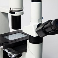
Shifting perspective from single microscope components to a full working live cell imaging solution, Leica Microsystems integrates microscope, LAS X imaging software, cameras, and dedicated third-party components into a complete live cell imaging system.
Imaging Neurons: A Laboratory Manual. Imaging Neurons: A Laboratory Manual Columbia Univ., New York City, NY. Reviews and disseminates the most important techniques currently available for studying the structure and function of living cells with optical methods. For students and researchers in neuroscience, cell biology, and developmental
The second edition of Live Cell Imaging: A Laboratory Manual expands upon and extends the collection of established and evolving methods for studying dynamic changes in living cells and organisms presented in the well-known first edition. There are 16 new …
Although presented primarily as a laboratory manual, the book includes introductory and background material and could be used as a textbook in advanced courses. It also includes a CD containing movies of living cells in action, created by investigators using the imaging techniques discussed in …
Download citation. Share . Facebook. Twitter. LinkedIn. Reddit. Request full-text. Live Cell Imaging: A Laboratory Manual. Article in Journal of Biomedical Optics 11(1):19901 · January 2006 with
The Journal of Biomedical Optics (JBO) is an open access journal that publishes peer-reviewed papers on the use of novel optical systems and techniques for improved health care and biomedical research.
Next Generation Live Cell Imaging System. Laser for imaging Laser for stimulation 1 Simultaneous Laser Light Stimulation and Imaging The FV1000 incorporates 2 laser scanners in a single compact design for simultaneous confocal fluorescence observation and independent laser light stimulation. Synchronization of these two functions ensures that cellular reactions that occur during or immediately
15/01/2011 · In this article and the accompanying poster, we will describe some of the general properties of FPs that are important to their function. We will also provide examples of successful mutagenesis that has been used to improve the use of these proteins for live-cell imaging…
Live Cell Imaging Tips . Imaging live cells is a complex task even for experienced microscopists. However, many research questions can only be addresed in living cells. For example studies of cellular dynamics and cell physiology require live observation over time. Additionally, microscopy of GFP-labeled proteins in living cells is both simpler and less prone to artifacts than microscopy of
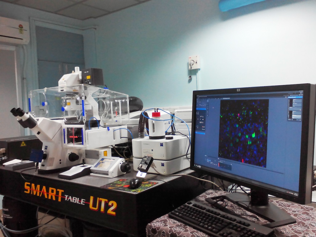
These results clearly demonstrate that the portable long-term live-cell imaging system is suitable to biomedical research. 3. Discussion. To overcome the limitations of commercially available technology suitable for long-term live-cell imaging we have developed a platform that is advantageous for several reasons. First, with a footprint of 22 – live cell imaging a laboratory manual
Live Cell Imaging A Laboratory Manual Robert D. Goldman
Book Review Live Cell Imaging A Laboratory Manual
![]()
Live-Cell Imaging MicroscopyU
IncuCyte® Live Cell Analysis System Sartorius

Live Cell Imaging A Laboratory Manual NASA/ADS
Image Analysis Software PerkinElmer
Home IPRASENSE
Imaging Neurons A Laboratory Manual
– Live Cell Imaging Indiana University
Nanolive SA > Looking inside life
Live Cell Imaging Reagents Sigma-Aldrich
Imaging A Laboratory Manual aetatrict.dustmasters.co.uk
IncuCyte® Live-Cell Analysis Systems are real-time quantitative live-cell imaging and analysis platforms that enable visualization and quantification of cell behavior over time, by automatically gathering and analyzing images around the clock within a standard laboratory incubator. This enables researchers to make time-lapsed, kinetic
Live-cell microscopy – tips and tools Journal of Cell
Live Cell Imaging A Laboratory Manual CSHL Scientific