Physics of ultrasound imaging pdf
History of Ultrasound and Technological Advances Jim Tsung, MD, MPH New York, USA. 3 Themes •Asking a question •Making an observation •Solving a problem. 1794: Lazzaro Spallanzani – Physiologist First to Study Ultrasound Physics by Deducing Bats used to ultrasound to navigate by echolocation. 1826: Jean Daniel Colladon –Physicist Uses Under-Water Church Bell (early ultrasound
Physics of Ultrasound Imaging. William Tod Drost. Ultrasound examinations are a widely used, indispensable diagnostic imaging test. It is a highly user-dependant interaction among the sonographer, patient, and machine.
Ultrasound Imaging Yao Wang Polytechnic University, Brooklyn, NY 11201 Based on J. L. Prince and J. M. Links, Medical Imaging Signals and Systems, and lecture notes by Prince.
The use of ultrasound imaging was found to be associated with significantly less risk of arterial puncture and haematomas and less time to insert the catheter as well as a higher success rate for inserting the catheter on the first attempt.
Understanding the basic ultrasound physics presented in this chapter will be helpful for anesthesiologists to appropriately select the transducer, to set the ultrasound system, and then to obtain a pleasing imaging
Basic Physics of Ultrasound – echolocation used by bats, whales and dolphins, as well The image is a 2D map of reflections displayed as a grey scale. B mode = brightness modulation ‘ The image is a 2D map of reflections displayed as a grey scale.
Ultrasound imaging utilizes the interaction of sound waves with living tissue to produce an image of the tissue or, in Doppler-based modes, determine the velocity of a moving tissue, primarily blood.
many areas, ultrasound is now chosen as the first line of investigation, before alternative imaging techniques. This book describes the physics and technology
The appearance of ultrasound images depends critically on the physical interactions of sound with the tissues in the body. The basic principles of ultrasound imaging and the physical reasons for many common artifacts are described.
The Essential Physics of Medical Imaging Pdf mediafire.com, rapidgator.net, 4shared.com, uploading.com, uploaded.net Download Note: If you’re looking for a free download links of The Essential Physics of Medical Imaging Pdf, epub, docx and torrent then this site is not for you.
Ultrasound is a form of non-ionizing radiation that uses high-frequency sound waves to image the body. It is a real-time investigation which allows assessment of moving structures and also facilitates measurement of velocity and direction of blood flow within a vessel.
physics of ultrasonic imaging. Knowledge of the basic physics of ultrasound is essential as a foundation for the understanding of the nature and behaviour of ultrasound, the mechanisms by which it
29/05/2011 · BASIC PHYSICS. Medical ultrasound machines generate ultrasound waves and receive the reflected echoes. Brightness mode (B mode) is the basic mode that is usually used. The B mode gives a two dimensional (2D) black and white image that depends on the anatomical site of the slice.
LEARNING OBJECTIVES 1) Relate the physical principles of traditional and new ultrasound imaging modes and processing to selection and application of these modes, including compound imaging
Lectures by Modality; Study Resources ; Contact Us . The content within this website is solely for educational purposes. For questions or comments, please contact the faculty listed on the Contact Us page. Lectures by Modality. CURRENTLY UNDER CONSTRUCTION! Quick Navigation * Radiography & Mammography * Fluoroscopy * Computed Tomography * Ultrasound * Magnetic Resonance Imaging …
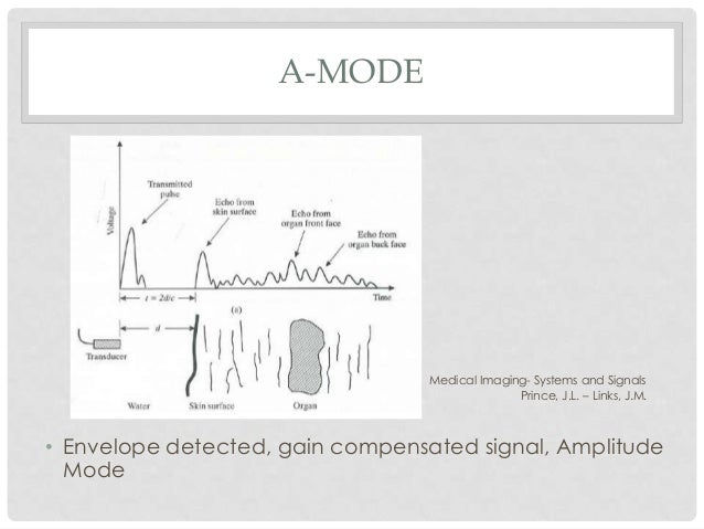
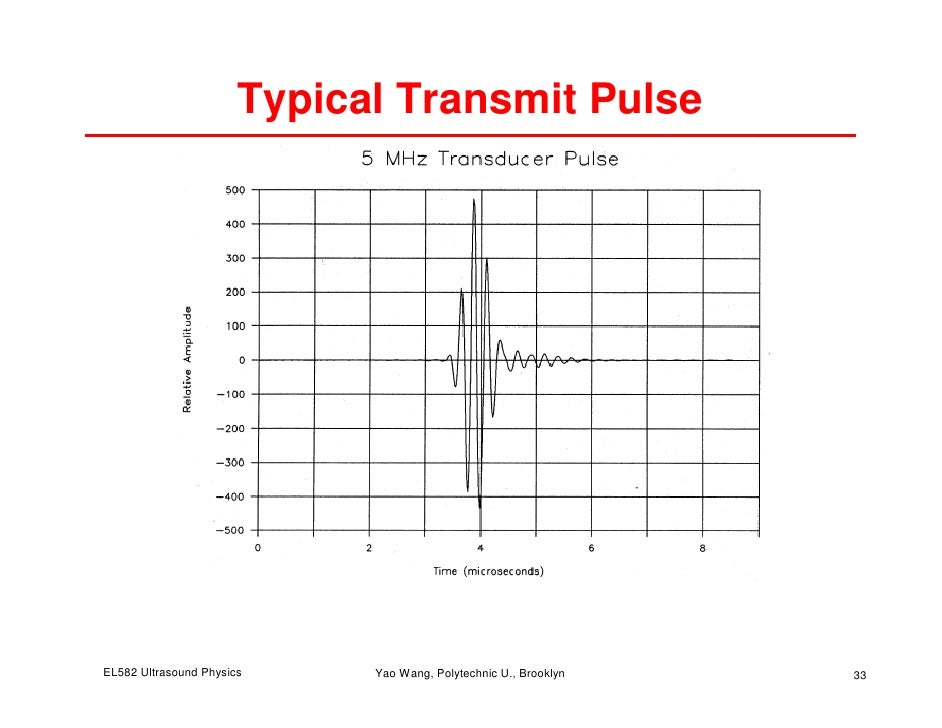
The Physics of Echo! Imaging! – Electrical stimulate piezoelectric crystal which sends ultrasound pulse !! – Transducer then “listens” for returning ultrasound signals! – Transducer “listens” 99 percent of time, which increases sensitivity! The Physics of Echo! 1-2 µsec! 0.4 µsec! 9/30/13! 7! Modes:! • A Mode – amplitude mode. Where the signals are displayed as spikes that
Physics, Instrumentation and New Trends. in Techniques of Ultrasound Medical Imaging Imaging: after energy (light, radio waves, Ultrasound and more) is interacting with a
PPT – Basic Ultrasound Physics PowerPoint presentation
Download The Essential Physics of Medical Imaging Pdf Ebook
Ultrasound or ultrasonography is a medical imaging technique that uses high frequency sound waves and their echoes. The technique is similar to the echolocation used by bats, whales and dolphins, as well as SONAR used by submarines .
Introduction to Physics and Applications of Vascular Imaging David E. Hintenlang, Ph.D. DABR, FACMP University of Florida Gainesville, FL ACMP 2009 Annual Meeting
Edelman Ultrasound Physics · 1 chapter 1 Ultrasound Physics Sidney K. Edelman, Ph.D. ESP Ultrasound www.esp-inc.com edelman@esp-inc.com Definitions Sound creates images by sending short bursts into the body.
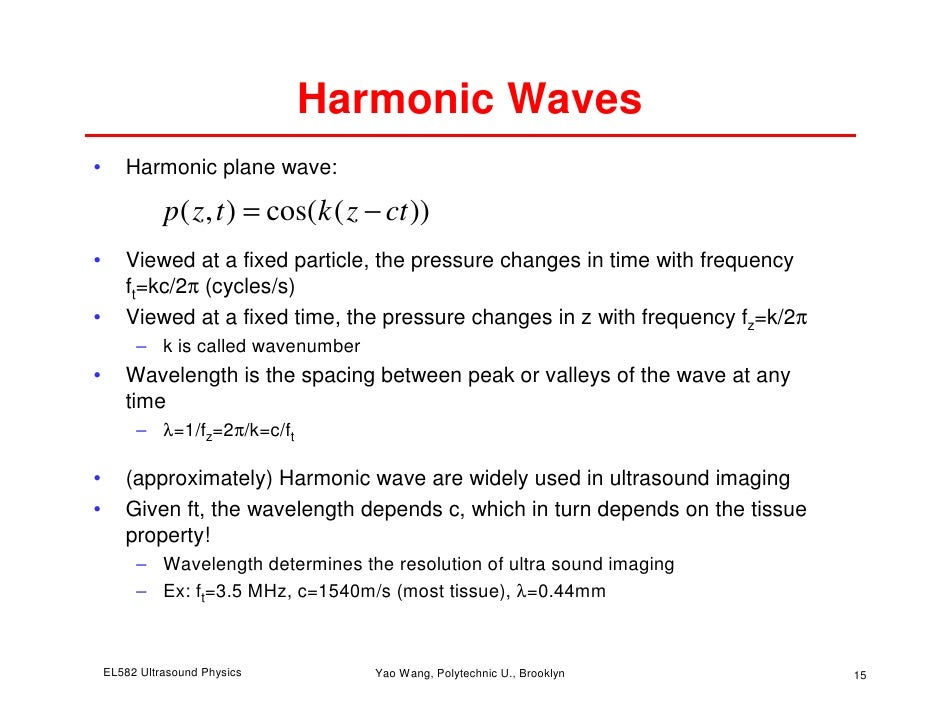
Ultrasound transducers contain a range of ultrasound frequencies, termed bandwidth. For example, 2.5-3.5 MHz for general abdominal imaging and 5.0-7.5 MHz for superficial imaging. For example, 2.5-3.5 MHz for general abdominal imaging and 5.0-7.5 MHz for superficial imaging.
Physics of Ultrasound UltrasoundImaging and Artifacts รศ.นพ.เดโช จกราพานั ิชกุล สาขาหทยวัทยาิ, ภาควชาอายิ ุรศาสตร ์คณะแพทยศาสตร์ศริราชพยาบาลิ
HSC Physics Notes – Medical Physics 9.4 – 1. The properties of ultrasound waves can be used as diagnostic tools 1. identify the differences between ultrasound and sound in normal hearing range Ultrasound is very high frequency sound. Ultrasound waves are sound waves with frequency greater than that of normal human hearing. That is, the frequency greater than → 20 000 hertz. • Sound

Beginning with basic physics of ultrasound, in the presentation how an ultrasound image is constructed is tried to be revealed by investigation of the wave propagation through the tissue.
Preface: Covering the basics of X-rays, CT, PET, nuclear medicine, ultrasound, and MRI, this textbook provides senior undergraduate and beginning graduate students with a broad introduction to medical imaging.
Download chapter PDF. The increasing availability of inexpensive portable ultrasound systems with a wide range of hardware and software options has allowed their widespread use in urology. To optimise the image, increase the diagnostic confidence of the operator, and minimise interpretive errors, it is necessary to understand the basic physics and technology underpinning these systems
2 Physics of Ultrasound Notes Lynette Hassall DMU AMS MLI, Clinical Applications Specialist, SonoSite, Inc. These notes are not a complete physics text, vast amounts of possibly significant information have been omitted to try to keep
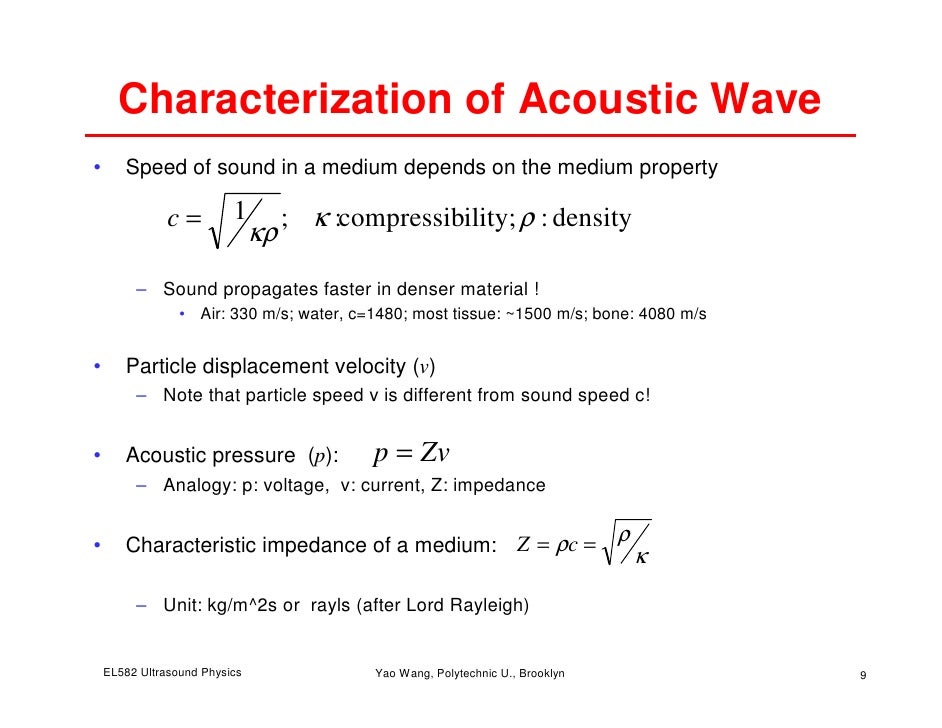
Ultrasound used in medical imaging typically operate at frequencies way above human hearing: about 2 million Hz to 20 million Hz (2-20 MHz). Generation of Ultrasound Waves To use ultrasound to find things, we first need to have a way of generating them.

• describe and compare processes of, and images produced by, medical imaging using two or more of ultrasound, X-rays, CT, MRI and PET; • identify and apply …
CHAPTER 1 • Basic physics of medical ultrasound. 4. average speed of 1540 m/s for conversion of time into depth. The high speed in bone can cause severe problems as will be seen later
Introduction to Medical Imaging Physics, Engineering and Clinical Applications Covering the basics of X-rays, CT, PET, nuclear medicine, ultrasound and MRI, this
Ultrasound is a form of non-ionizing radiation that uses high-frequency sound waves to image the body. It is a real-time investigation which allows assessment of moving structures and also facilitates measurement of velocity and directionality of blood flow within a vessel.
Learning the physics of medical imaging is a continuing and progressive process that uses a series of learning activities as illustrated here. Each activity has its values and limitations. Each activity has its values and limitations.
UW Imaging Physics Course University of Washington
Ultrasound imaging systems uses piezoelectric transducers as source and detector. Piezoelectric crystals vibrate in response to an alternating voltage, and when placed against a patient’s skin and driven at high frequencies produce ultrasound pulses that travel
Ultrasound contrast agents are also used for the measurement of flow by Doppler-imaging or related techniques. Therefore, a short summary of ultrasound flow measurement techniques will be given. Application examples for anatomical and molecular imaging with microbubble contrast media will be presented and discussed.
If an ultrasound system is used for imaging, it must use pulsed ultrasound and, therefor e, the duty factor must be between 0% and 100% (or 0 and 1), typically close to 0.
Anaesthesia Tutorial of the Week 218 – The Physics of Ultrasound: Part 2, 21/03/11 Page 6 of 8 As the transmitted frequency, speed of sound in tissue and Doppler shift is known, this equation can be
Physics of Ultrasound Imaging Yao Wang Polytechnic University, Brooklyn, NY 11201 Based on J. L. Prince and J. M. Links, Medical Imaging Signals and Systems, and lecture notes by Prince. Figures are from the textbook except otherwise noted.
This comprehensive publication covers all aspects of image formation in modern medical imaging modalities, from radiography, fluoroscopy, and computed tomography, to magnetic resonance imaging and ultrasound.
Basic physics of ultrasound imaging John E. Aldrich, PhD, FCCPM T o accurately interpret ultra-sound images, a basic under-standing of the physical prin-ciples involved in ultrasound image generation is essential. Although often considered a simple bedside tech-nology, these principles can be somewhat complicated. A comparison with those pertaining to radiographic imaging illus-trates this
The Physics of Radiology and Imaging PDF Preface Publisher’ Note: Products purchased from 3rd Party sellers are not guaranteed by the Publisher for quality, authenticity, or access to any online entitlements included with the product. – principles of magnetic resonance imaging dwight nishimura pdf Ultrasound imaging or sonography is often used in medicine. In the nondestructive testing of products and structures, ultrasound is used to detect invisible flaws. Industrially, ultrasound is used for cleaning, mixing, and accelerating chemical processes.
imaging, where the emitter (the X-ray tube) and recorder (the detectors) are located on the opposite side of the patient. This document attempts to give simple insight in to basic ultrasound…
Ultrasound produces an image, and how to optimize those images to achieve the best from your machine, for the benefit of your patients. Please refer to Physics text books and articles for more complete explanations.
Physics of Ultrasound; Echocardiography (FATE) SEARCH. Ultrasound Workshops for 2019. Focus Assessed Transthoracic Echocardiography (FATE) Workshop. FATE is Focus Assessed Transthoracic Echo. It is a one-day echocardiography workshop for physicians. FATE teaches delegates how to perform a basic echo study and provides the skills to interpret findings and place them in the clinical …
Page 121. Chapter 7 Ultrasonics 7.1 Introduction. Ultrasound, as currently practiced in medicine, is a real-time tomographic imaging modality. Not only does it produce real-time tomograms of scattering, but it can also be used to produce real-time images of tissue and blood motion, elasticity, and flow in the tissue (perfusion).
Anyone with an interest in clinical ultrasound imaging would benefit from this session. Page 2 of 8 Understanding and Teaching Ultrasound Physics Randell L. Kruger, Ph.D. INTRODUCTION There are ten clinical ultrasound physics demonstrations discussed in this course, participants are introduced to procedures used to demonstrate and explore the physics associated with these demonstrations
Today’s Topics ! History ! What is Ultrasound? ! Physics of ultrasound ! Ultrasonic echo imaging ! Focusing technique ! A-mode signal and B-mode image
To familiarize students with Physics or Ultrasound, commonly used in diagnostic imaging modality. Chapter 12:Physics of Ultrasound Slide set prepared by E.Okuno (S. Paulo, Brazil, Institute of Physics of S. Paulo University) IAEA 12.1. Introduction 12.2. Ultrasonic Plane Waves 12.3. Ultrasonic Properties of Biological Tissue 12.4. Ultrasonic Transduction 12.5. Doppler Physics 12.6. Biological
Kollmann C. (2004) Basic Principles and Physics of Duplex and Color Doppler Imaging. In: Mostbeck G.H. (eds) Duplex and Color Doppler Imaging of the Venous System. Medical Radiology (Diagnostic Imaging and Radiation Oncology). Springer, Berlin, Heidelberg
ATOTW 199 The physics of ultrasound – part 1 04/10/2010 Page 2 of 8 BASIC PRINCIPLES OF SOUND WAVES Sound is mechanical energy that is transmitted through …
BATS Better Anaesthesia Through Sonography
Physics of Ultrasound NYSORA
Basic physics of ultrasound imaging Critical Care Medicine
How Ultrasound Works Department of Physics
Introduction For physics
Clinical ultrasound physics PubMed Central (PMC)
X-ray imaging Institute of Physics – For physics
The Physics of Ultrasound SpringerLink
– Mathematics and Physics of Emerging Biomedical Imaging
Physics of Ultrasound Imaging Veterian Key
Physics of Breast Ultrasound Request PDF ResearchGate
199 The physics of ultrasound part 1 – AAGBI
This comprehensive publication covers all aspects of image formation in modern medical imaging modalities, from radiography, fluoroscopy, and computed tomography, to magnetic resonance imaging and ultrasound.
Reflection-mode ultrasound imaging University of Michigan
• describe and compare processes of, and images produced by, medical imaging using two or more of ultrasound, X-rays, CT, MRI and PET; • identify and apply …
The Physics of Ultrasound Request PDF ResearchGate
Ultrasound imaging ch11 New York University Tandon
Physics of ultrasound imaging SlideShare
Basic Physics of Ultrasound – echolocation used by bats, whales and dolphins, as well The image is a 2D map of reflections displayed as a grey scale. B mode = brightness modulation ‘ The image is a 2D map of reflections displayed as a grey scale.
Basic Principles and Physics of Duplex and Color Doppler
Physics of Ultrasound NYSORA