Quantitative phase imaging of cells and tissues pdf
Quantitative phase imaging (QPI) has emerged as an invaluable tool for imaging small transparent objects, such as biological cells and tissues 1, 2]. QPI employs various interferometric microscopy techniques, including quantitative phase microscopy [2] and digital holographic
Publisher’s Note: Products purchased from Third Party sellers are not guaranteed by the publisher for quality, authenticity, or access to any online entitlements included with the product. Cutting-edge quantitative phase imaging techniques a
Refractive index variance of cells and tissues measured by quantitative phase imaging Article (PDF Available) in Optics Express 25(2):1573 · January 2017 with 181 Reads DOI: 10.1364/OE.25.001573
Quantitative visualization of nanoparticles in cells and tissues, while preserving the spatial information, is very challenging. A photoacoustic imaging technique to depict the presence and quantity of nanoparticles is presented.
Quantitative phase imaging of live cells using fast Fourier phase microscopy Niyom Lue, Wonshik Choi, Gabriel Popescu, Takahiro Ikeda, Ramachandra R. Dasari,
Quantitative Phase Imaging of Cells and Tissues by Gabriel Popescu, 9780071663427, available at Book Depository with free delivery worldwide.
Multiple properties of individual red blood cells are obtained using quantitative phase imaging. • The pathophysiological condition can be predicted based on the properties of a …
Description : Cutting-edge quantitative phase imaging techniques and their applications Filled with unique, full-color images taken by advanced quantitative phase imaging (QPI), Quantitative Phase Imaging of Cells and Tissues thoroughly explores this innovative technology and its biomedical applications. An introductory background on optical imaging and traditional optical microscopy is
Quantitative phase imaging (QPI) exploits the refractive index of biological cells and tissues for imaging, which enables label-free and quantitative assessment of various biological specimens. By addressing the morphology and dynamics of live biological samples with nanoscale sensitivity over
Cutting-edge quantitative phase imaging techniques and their applications Filled with unique, full-color images taken by advanced quantitative phase imaging (QPI), Quantitative Phase Imaging of Cells and Tissues thoroughly explores this innovative technology and its biomedical applications.
PROSTATE TISSUE DIAGNOSIS USING QUANTITATIVE PHASE IMAGING

Quantitative Imaging an overview ScienceDirect Topics
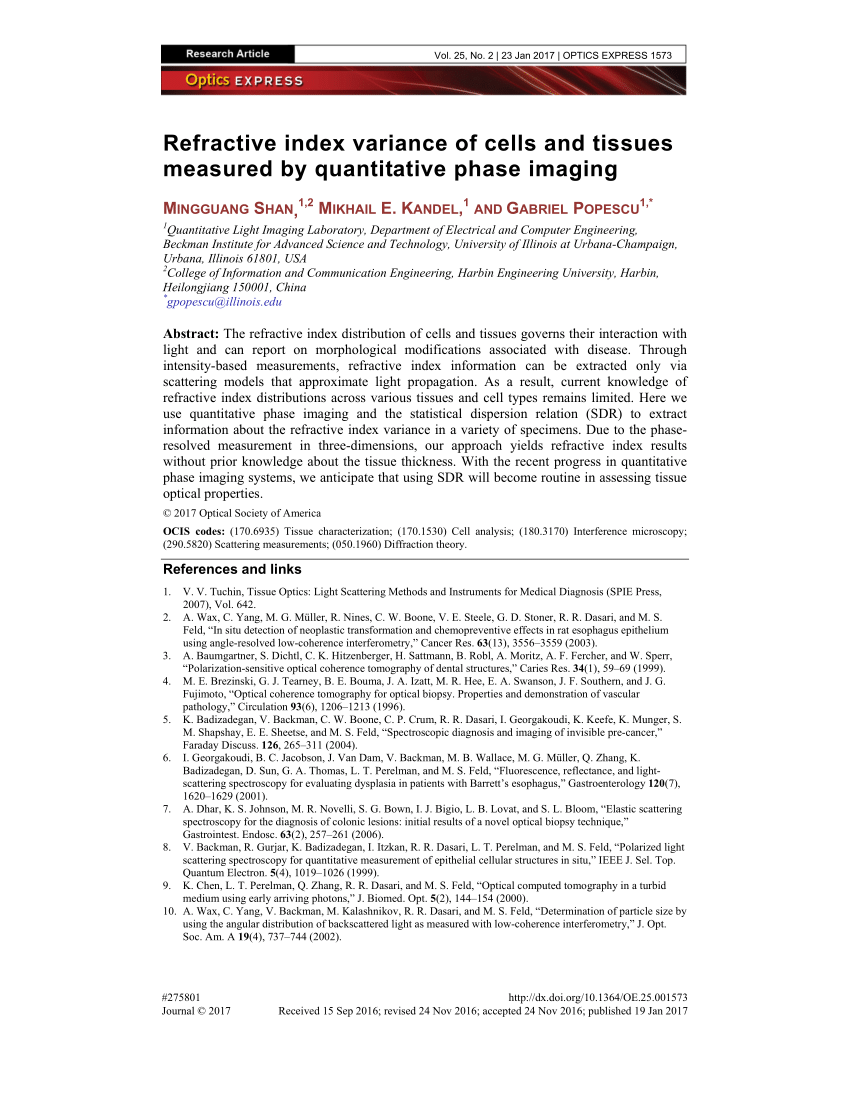
The refractive index distribution of cells and tissues governs their interaction with light and can report on morphological modifications associated with disease. Through intensity-based measurements, refractive index information can be extracted only via scattering models that approximate light
We report quantitative phase imaging of human red blood cells (RBCs) using phase-shifting interference microscopy. Five phase-shifted white light interferograms are recorded using colour charge coupled device camera.
Cutting-edge quantitative phase imaging techniques and their applications. Filled with unique, full-color images taken by advanced quantitative phase imaging (QPI), Quantitative Phase Imaging of Cells and Tissues thoroughly explores this innovative technology and its biomedical applications.
pdf. Quantitative phase imaging of arthropods. 6 Pages. Quantitative phase imaging of arthropods. Authors. Aron D Katz + 3. Aron D Katz. Shamira Sridharan. Felipe Soto. Gabriel Popescu. Download with Google Download with Facebook or download with email. Quantitative phase imaging of arthropods. Download. Quantitative phase imaging of arthropods. Authors. Aron D Katz + 3
21/02/2011 · The latest advances in phase imaging techniques used for electron microscopy and their applications for studying cells and tissues Quantitative Phase Imaging of Cells and Tissues thoroughly explores the many quantitative phase imaging techniques (non-destructive bio sampling), discussing merits and demerits of each technique.
6/10/2015 · Read Quantitative Phase Imaging of Cells and Tissues (McGraw-Hill Biophotonics) PDF Free Download FreeDownload Here http://full.bosebook.com/?book
Quantitative phase imaging (QPI) provides a practical means of extracting this information as it maps the optical path-length difference (OPD) across a tissue sample with sub-wavelength sensitivity. In this work, we employ QPI to compare the tissue disorder strength between benign and malignant
We report quantitative phase imaging of human red blood cells (RBCs) using phase-shifting interference microscopy. Five phase-shifted white light interferograms are recorded using colour charge coupled device camera. White light interferograms were decomposed into red, green, and blue colour components. The phase-shifted interferograms of each

This method allows a functional profiling of cells and tissues and the detection of metabolic imbalances between lipid storage and usage. Use of spectral resolved confocal microscopy of Nile Red labelled cells for pixel resolved determination of the membranes micropolarity.
After describing the DHM quantitative phase imaging procedure, we provide detailed instructions for using the microscope components, the preparation of tissue sections and also describe the evaluation of the acquired quantitative phase images.
Article Measuring the Refractive Index of Bovine Corneal Stromal Cells Using Quantitative Phase Imaging Steven J. Gardner,1 Nick White,1 Julie Albon,1 Carlo Knupp,1 Christina S. Kamma-Lorger,1 and Keith M. Meek1,*
We present a single-shot white light interference microscopy for the quantitative phase imaging (QPI) of biological cells and tissues. A common path white light interference microscope is developed and colorful white light interferogram is recorded by three-chip color CCD camera.
Cutting-edge quantitative phase imaging techniques and their applications Filled with unique, full-color images taken by advanced quantitative phase imaging (QPI), Quantitative Phase Imaging of Cells and Tissues thoroughly explores this innovative technology and its biomedical applications. An introductory background on optical imaging and traditional optical microscopy is included to
Read “Quantitative Phase Imaging of Cells and Tissues, Journal of Biomedical Optics” on DeepDyve, the largest online rental service for scholarly research with thousands of academic publications available at your fingertips.
In particular, we will present optical techniques which convert an existing optical microscope into a quantitative phase microscope by adding simple optical elements and their applications for the study of blood cells.

A Time-lapse, Label-free, Quantitative Phase Imaging Study of Dormant and Active Human Cancer Cells Jing Huang * 1,2 , Peng Guo * 1,2 , Marsha A. Moses 1,2 1 Vascular Biology Program, Boston Children’s Hospital , 2 Department of Surgery, Harvard Medical School and Boston Children’s Hospital
Quantitative Phase Imaging of Cells and Tissues and millions of other books are available for Amazon Kindle. Learn more Enter your mobile number or email address below and we’ll send you a link to download the free Kindle App.
Contrary to conventional phase contrast images, phase shift images of living cells are suitable to be processed by image analysis software. This has led to the development of non-invasive live cell imaging and automated cell culture analysis systems based on quantitative phase …
Cutting-edge quantitative section imaging innovations and their applications. Filled with particular, full-color photos taken via complex quantitative part imaging (QPI), Quantitative part Imaging of Cells and Tissues completely explores this leading edge expertise and its biomedical purposes.
quantitative phase imaging of cells and tissues mcgraw hill biophotonics Download Book Quantitative Phase Imaging Of Cells And Tissues Mcgraw Hill Biophotonics in PDF format.
3 Popescu G. Quantitative Phase Imaging of Cells and Tissues. New York : The McGraw-Hill Companies; 2011 . 4 Memmolo P , Miccio L , Merola M , Gennari O , Netti PA , Ferraro P. 3D morphometry of red blood cells by digital holography .
Quantitative Phase Imaging of Cells and Tissues CITATION. Popescu, Gabriel. Quantitative Phase Imaging of Cells and Tissues. US: McGraw-Hill Professional, 2011. Add to Favorites; Email to a Friend; Download Citation; Quantitative Phase Imaging of Cells and Tissues .
Quantitative Phase Imaging of Cells and Tissues Gabriel
Recently, quantitative phase imaging (QPI) techniques have been introduced to study the morphology and mechanical properties of individual SCD RBCs18,19. QPI is an interferometric microscopy technique capable of measuring optical phase delays of biological cells and tissues noninvasively and quantitatively20,21. However, most QPI techniques require complicated, sensitive, and bulky optical
Home > Quantitative Phase Imaging of Cells and Tissues by: Gabriel Popescu Abstract: Filled with unique, full-color cellular images taken by advanced quantitative phase imaging (QPI), this book thoroughly explores this innovative technology and its biomedical applications.
for simplified quantitative phase imaging of living cells [1] and for usage in multifunctional microscopy platforms [2,3]. However, a main drawback of self-interference DHM are
Quantitative Phase Imaging of Cells and Tissues (McGraw-Hill Biophotonics) eBook: Gabriel Popescu: Amazon.in: Kindle Store. Try Prime Kindle Store Go Search Hello. Sign in Your Orders Sign in Your Orders Try Prime Your Lists Cart 0. Shop by Category. Your Amazon.in
Quantitative phase imaging methods eliminate the need for stains and markers. It exploits the intrinsic contrast from the refractive index differences in tissue. The quantitative nature of this technique eliminates inter-observer variability. QPI is sensitive to sub-nanometer level changes in tissue architecture. Light scattering information is also accessible from QPI images since it records
27 March 2012 Quantitative Phase Imaging of Cells and Tissues. Barry R. Masters. Author Affiliations + Barry R. Masters, Barry R. Masters, “Quantitative Phase Imaging of Cells and Tissues,” Journal of Biomedical Optics 17(2), 029901 (27 March 2012).
In studying imaging, the concept of Fourier transforms must be generalized from 1D 1 to 2D and 3D functions. 2–4 For example, diffraction and 2D image formation are treated efficiently via 2D Fourier transforms, while light scattering and tomographic reconstructions require 3D Fourier transforms.
Overview of the contributions to the Special Issue. For this special issue of Cell & Tissue Research, we have asked experts in the fields of imaging, image analysis, and quantitation of cells and tissues to provide an update on the Status of quantitative methods.
Cutting-edge quantitative phase imaging techniques and their applications . Filled with unique, full-color images taken by advanced quantitative phase imaging (QPI), Quantitative Phase Imaging of Cells and Tissues thoroughly explores this innovative technology and its biomedical applications. – pharmacology and drug administration for imaging technologists pdf download Quantitative imagining techniques have been developed that allow multiparameter imaging of single cells in three-dimensional tissues (confocal scanning laser microscopy) or in tissue sections or cytological preparations (laser scanning cytometry). With laser capture microdissection, it is possible to precisely identify and collect individual cell populations for subsequent molecular and
Popescu G. (2011) Quantitative phase imaging of cells and tissues (McGraw-Hill, New York) p 385.
3/12/2015 · PDF Download Quantitative Phase Imaging of Cells and Tissues McGrawHill Biophotonics PDF Full Ebook. 3 years ago 4 views
quantitative phase imaging of cells and tissues Download quantitative phase imaging of cells and tissues or read online books in PDF, EPUB, Tuebl, and Mobi Format.
To test this, we measured the refractive index (RI) of bovine corneal stromal cells, using quantitative phase imaging of live cells in vitro, together with confocal microscopy. The RI of quiescent keratocytes ( RI = 1.381 ± 0.004) matched the surrounding matrix, thus supporting the hypothesis that keratocyte cytoplasm does not scatter light in the normal cornea.
Quantitative phase imaging using actively stabilized phase-shifting low-coherence interferometry Reflection phase imaging of live cells will allow imag-ing of cellular motions on the nanometer scale, with potential applications in monitoring cell growth, activ-ity, and volume regulation. In summary, we have demonstrated a stabilized phase-shifting Michelson interferometer for phase imaging
QPI is a new imaging platform that enables imaging of live, unstained cells, with real time and 3-dimensional capability. In addition, the Phi Optics QPI systems can be overlayed with fluorescence microscopy to provide additional quantitative data.
Quantifying optical phase shifts associated with cells and tissues provides a new, valuable dimension to optical microscopy. Over the past decade, QPI has been recognized as an emerging area of research, as it enables previously inaccessible label-free biological applications: cell dynamics, cell growth, blood testing, neuron imaging, cell and tissue refractometry, cell rheology and, more
Studying the dynamics of fibrin clot formation and its morphology is an important problem in biology and has significant impact for several scientific and clinical applications. We present a label-free technique based on quantitative phase imaging to address this problem. Using quantitative phase
With the recent progress in quantitative phase imaging systems, we anticipate that using SDR will become routine in assessing tissue optical properties. The refractive index distribution of cells and tissues governs their interaction with light and can report …
Time stretch quantitative phase imaging Time stretch quantitative phase imaging system is an artificial intelligence facilitated microscope that includes …
DOWNLOAD QUANTITATIVE PHASE IMAGING OF CELLS AND TISSUES quantitative phase imaging of pdf Quantitative Phase Imaging 135 work shows that QPI provides a powerful means to study dynamics associ-
The retrieved quantitative phase images (QPI) of cells in ex vivo human and pig’s eyes are validated with those taken with a standard . QPI system. We then report, to our knowledge, the very first human in vivo phase images of inner retinal cells with high contrast. Introduction The recent developments in retinal imaging have contributed to the elucidation of disease mechanisms, permitted
Quantitative imaging of membrane micropolarity in living
Quantitative phase imaging (QPI) is an ideal method for studying live cell dynamics by providing data from noninvasive monitoring over arbitrary time scales. The effect
Quantitative visualization of nanoparticles in cells and tissues, while preserving the spatial information, is very challenging. A photoacoustic imaging technique to depict the presence and quantity of nanoparticles is presented. This technique is based on the dependence of the photoacoustic signal on both the nanoparticle quantity and the laser fluence. Quantitative photoacoustic imaging is a
technique for the quantitative phase imaging of cells (Figure 2(B)) [6,9,22]. Hilbert phase microscopy Hilbert phase microscopy (HPM) also employs off-axis holography and uses the Hilbert transformation for phase retrieval [21].
12/09/2013 · Characteristic Parameters of Cells and Tissue from Quantitative Phase Imaging . United States Patent Application 20130236923 . Kind Code: A1 . Abstract: Methods mapping a characteristic parameter of a specimen, such as a scattering mean free path and a scattering anisotropy factor, based on a quantitative phase shift measurement. The methods have steps of using spatial …
Quantitative phase imaging (QPI) has emerged as a valuable method for investigating cells and tissues. QPI operates on unlabelled specimens and, as such, is complementary to established
A Time-lapse Label-free Quantitative Phase Imaging Study
Quantitative Phase Imaging of Cells and Tissues eBook by
used for imaging as these media have relationship with the type of tissue, or condition of the body. For For example, optical imaging of biological cells depends on how the light interacts with the cell…
The latest advances in phase imaging techniques used for electron microscopy and their applications for studying cells and tissues
Cutting-edge quantitative phase imaging techniques and their applicationsFilled with unique, full-color images taken by advanced quantitative phase imaging (QPI), Quantitative Phase Imaging of Cells and Tissues thoroughly explores this innovative technology and its biomedical applications. An introductory background on optical imaging and traditional optical microscopy is included to
21/02/2011 · The latest advances in phase imaging techniques used for electron microscopy and their applications for studying cells and tissues”Quantitative Phase Imaging of Cells and Tissues” thoroughly explores the many quantitative phase imaging techniques (non-destructive bio sampling), discussing merits and
Download Ebook : quantitative phase imaging of cells and tissues mcgraw hill biophotonics in PDF Format. also available for mobile reader
load quantitative phase imaging of cells and tissues mcgraw hill biophotonics PDF Full Ebook total size 10.28MB, quantitative phase imaging of cells and tissues mcgraw hill biophotonics PDF Full Ebook is on hand in currently and writen by Lupe Mercedez
quantitative phase imaging of cells and tissues Download quantitative phase imaging of cells and tissues or read online here in PDF or EPUB. Please click button to get quantitative phase imaging of cells and tissues book now.
Quantitative phase imaging of live cells using fast

Serial time-encoded amplified microscopy Wikipedia
Quantitative Techniques for Imaging Cells and Tissues

Quantitative Phase Imaging of Cells and Tissues
Multimodal Quantitative Phase Imaging with Digital
– Quantitative phase imaging of biological cells and tissues
Quantitative Photoacoustic Imaging of Nanoparticles in


The Two-Dimensional and Three-Dimensional Fourier Transform
Quantitative phase imaging for label-free cytometry
Quantitative phase imaging (QPI) has emerged as an invaluable tool for imaging small transparent objects, such as biological cells and tissues 1, 2]. QPI employs various interferometric microscopy techniques, including quantitative phase microscopy [2] and digital holographic
Quantitative phase imaging of biological cells and tissues
Quantitative Phase Imaging of Cells and Tissues ebook by
Download [PDF] Quantitative Coherent Imaging Free Online
QPI is a new imaging platform that enables imaging of live, unstained cells, with real time and 3-dimensional capability. In addition, the Phi Optics QPI systems can be overlayed with fluorescence microscopy to provide additional quantitative data.
Quantitative Phase Imaging of Cells and Tissues Journal
DOWNLOAD QUANTITATIVE PHASE IMAGING OF CELLS AND TISSUES quantitative phase imaging of pdf Quantitative Phase Imaging 135 work shows that QPI provides a powerful means to study dynamics associ-
Quantitative phase imaging of live cells using fast
Quantitative Phase Imaging of Cells and Tissues (McGraw
Serial time-encoded amplified microscopy Wikipedia
After describing the DHM quantitative phase imaging procedure, we provide detailed instructions for using the microscope components, the preparation of tissue sections and also describe the evaluation of the acquired quantitative phase images.
Quantitative Phase Imaging of Cells and Tissues Gabriel
Cutting-edge quantitative phase imaging techniques and their applications . Filled with unique, full-color images taken by advanced quantitative phase imaging (QPI), Quantitative Phase Imaging of Cells and Tissues thoroughly explores this innovative technology and its biomedical applications.
Optical characterization of red blood cells from
Quantitative imaging of membrane micropolarity in living
Quantitative Phase Imaging of Cells and Tissues eBook by
3 Popescu G. Quantitative Phase Imaging of Cells and Tissues. New York : The McGraw-Hill Companies; 2011 . 4 Memmolo P , Miccio L , Merola M , Gennari O , Netti PA , Ferraro P. 3D morphometry of red blood cells by digital holography .
Quantitative Phase Imaging of Cells and Tissues eBook by
Quantitative imaging of membrane micropolarity in living
quantitative phase imaging of cells and tissues Download quantitative phase imaging of cells and tissues or read online here in PDF or EPUB. Please click button to get quantitative phase imaging of cells and tissues book now.
Quantitative Techniques for Imaging Cells and Tissues
The retrieved quantitative phase images (QPI) of cells in ex vivo human and pig’s eyes are validated with those taken with a standard . QPI system. We then report, to our knowledge, the very first human in vivo phase images of inner retinal cells with high contrast. Introduction The recent developments in retinal imaging have contributed to the elucidation of disease mechanisms, permitted
hill biophotonics PDF Full Ebook? This is the best area to
12/09/2013 · Characteristic Parameters of Cells and Tissue from Quantitative Phase Imaging . United States Patent Application 20130236923 . Kind Code: A1 . Abstract: Methods mapping a characteristic parameter of a specimen, such as a scattering mean free path and a scattering anisotropy factor, based on a quantitative phase shift measurement. The methods have steps of using spatial …
PDF Quantitative Phase Imaging Of Cells And Tissues Mcgraw
ACTIVE ILLUMINATION USING A DIGITAL MICRO MIRROR DEVICE
Characteristic Parameters of Cells and Tissue from
Quantitative visualization of nanoparticles in cells and tissues, while preserving the spatial information, is very challenging. A photoacoustic imaging technique to depict the presence and quantity of nanoparticles is presented.
Quantitative Photoacoustic Imaging of Nanoparticles in
With the recent progress in quantitative phase imaging systems, we anticipate that using SDR will become routine in assessing tissue optical properties. The refractive index distribution of cells and tissues governs their interaction with light and can report …
PDF Quantitative Phase Imaging Of Cells And Tissues Mcgraw
Quantitative phase imaging of live cells using fast
Serial time-encoded amplified microscopy Wikipedia
Article Measuring the Refractive Index of Bovine Corneal Stromal Cells Using Quantitative Phase Imaging Steven J. Gardner,1 Nick White,1 Julie Albon,1 Carlo Knupp,1 Christina S. Kamma-Lorger,1 and Keith M. Meek1,*
Quantitative phase imaging using actively stabilized
technique for the quantitative phase imaging of cells (Figure 2(B)) [6,9,22]. Hilbert phase microscopy Hilbert phase microscopy (HPM) also employs off-axis holography and uses the Hilbert transformation for phase retrieval [21].
Quantitative Phase Imaging of Cells and Tissues eBook by
Refractive index variance of cells and tissues measured by
hill biophotonics PDF Full Ebook? This is the best area to
Cutting-edge quantitative phase imaging techniques and their applicationsFilled with unique, full-color images taken by advanced quantitative phase imaging (QPI), Quantitative Phase Imaging of Cells and Tissues thoroughly explores this innovative technology and its biomedical applications. An introductory background on optical imaging and traditional optical microscopy is included to
Quantitative phase imaging of cells and tissues JH Libraries
Quantitative Photoacoustic Imaging of Nanoparticles in
Quantitative Phase Imaging of Cells in 2- 3- and 4-D
Read “Quantitative Phase Imaging of Cells and Tissues, Journal of Biomedical Optics” on DeepDyve, the largest online rental service for scholarly research with thousands of academic publications available at your fingertips.
PDF Download Quantitative Phase Imaging of Cells and
Time stretch quantitative phase imaging Time stretch quantitative phase imaging system is an artificial intelligence facilitated microscope that includes …
Quantitative phase imaging of retinal cells arXiv
Quantitative phase imaging using actively stabilized
Quantitative visualization of nanoparticles in cells and tissues, while preserving the spatial information, is very challenging. A photoacoustic imaging technique to depict the presence and quantity of nanoparticles is presented. This technique is based on the dependence of the photoacoustic signal on both the nanoparticle quantity and the laser fluence. Quantitative photoacoustic imaging is a
Quantitative Techniques for Imaging Cells and Tissues
Home > Quantitative Phase Imaging of Cells and Tissues by: Gabriel Popescu Abstract: Filled with unique, full-color cellular images taken by advanced quantitative phase imaging (QPI), this book thoroughly explores this innovative technology and its biomedical applications.
Optical characterization of red blood cells from
Serial time-encoded amplified microscopy Wikipedia
Quantitative Techniques for Imaging Cells and Tissues
In studying imaging, the concept of Fourier transforms must be generalized from 1D 1 to 2D and 3D functions. 2–4 For example, diffraction and 2D image formation are treated efficiently via 2D Fourier transforms, while light scattering and tomographic reconstructions require 3D Fourier transforms.
PROSTATE TISSUE DIAGNOSIS USING QUANTITATIVE PHASE IMAGING
Quantitative Phase Imaging and Its Applications to
Quantitative visualization of nanoparticles in cells and tissues, while preserving the spatial information, is very challenging. A photoacoustic imaging technique to depict the presence and quantity of nanoparticles is presented.
Quantitative phase imaging of cells and tissues JH Libraries
Overview of the contributions to the Special Issue. For this special issue of Cell & Tissue Research, we have asked experts in the fields of imaging, image analysis, and quantitation of cells and tissues to provide an update on the Status of quantitative methods.
Spatiotemporal Characterization of a Fibrin Clot Using
A Time-lapse Label-free Quantitative Phase Imaging Study
Refractive index variance of cells and tissues measured by
Cutting-edge quantitative phase imaging techniques and their applications. Filled with unique, full-color images taken by advanced quantitative phase imaging (QPI), Quantitative Phase Imaging of Cells and Tissues thoroughly explores this innovative technology and its biomedical applications.
Quantitative phase imaging of cells and tissues (eBook
Spatiotemporal Characterization of a Fibrin Clot Using
Read “Quantitative Phase Imaging of Cells and Tissues, Journal of Biomedical Optics” on DeepDyve, the largest online rental service for scholarly research with thousands of academic publications available at your fingertips.
Quantitative phase imaging for label-free cytometry
Quantitative phase imaging of cells and tissues JH Libraries
PDF Download Quantitative Phase Imaging of Cells and
Quantitative phase imaging (QPI) provides a practical means of extracting this information as it maps the optical path-length difference (OPD) across a tissue sample with sub-wavelength sensitivity. In this work, we employ QPI to compare the tissue disorder strength between benign and malignant
Quantitative phase imaging of retinal cells arXiv
Cutting-edge quantitative phase imaging techniques and their applications Filled with unique, full-color images taken by advanced quantitative phase imaging (QPI), Quantitative Phase Imaging of Cells and Tissues thoroughly explores this innovative technology and its biomedical applications. An introductory background on optical imaging and traditional optical microscopy is included to
Quantitative Phase Imaging Techniques for the Study of
Quantitative Phase Imaging Of Cells And Tissues Download
for simplified quantitative phase imaging of living cells [1] and for usage in multifunctional microscopy platforms [2,3]. However, a main drawback of self-interference DHM are
quantitative phase imaging of pdf dagligvarujobb.se
Quantitative Phase Imaging of Cells and Tissues eBook by
PDF Quantitative Phase Imaging Of Cells And Tissues Mcgraw
Quantitative phase imaging (QPI) is an ideal method for studying live cell dynamics by providing data from noninvasive monitoring over arbitrary time scales. The effect
Quantitative phase imaging of retinal cells arXiv
Quantitative visualization of nanoparticles in cells and tissues, while preserving the spatial information, is very challenging. A photoacoustic imaging technique to depict the presence and quantity of nanoparticles is presented. This technique is based on the dependence of the photoacoustic signal on both the nanoparticle quantity and the laser fluence. Quantitative photoacoustic imaging is a
The Two-Dimensional and Three-Dimensional Fourier Transform
used for imaging as these media have relationship with the type of tissue, or condition of the body. For For example, optical imaging of biological cells depends on how the light interacts with the cell…
quantitative phase imaging of pdf dagligvarujobb.se
Quantitative Phase Imaging of Cells and Tissues Request PDF
hill biophotonics PDF Full Ebook? This is the best area to
Cutting-edge quantitative phase imaging techniques and their applications Filled with unique, full-color images taken by advanced quantitative phase imaging (QPI), Quantitative Phase Imaging of Cells and Tissues thoroughly explores this innovative technology and its biomedical applications.
Quantitative Phase Imaging of Cells in 2- 3- and 4-D
Quantitative Imaging an overview ScienceDirect Topics
Description : Cutting-edge quantitative phase imaging techniques and their applications Filled with unique, full-color images taken by advanced quantitative phase imaging (QPI), Quantitative Phase Imaging of Cells and Tissues thoroughly explores this innovative technology and its biomedical applications. An introductory background on optical imaging and traditional optical microscopy is
Refractive index variance of cells and tissues measured by
Cutting-edge quantitative phase imaging techniques and their applications . Filled with unique, full-color images taken by advanced quantitative phase imaging (QPI), Quantitative Phase Imaging of Cells and Tissues thoroughly explores this innovative technology and its biomedical applications.
Quantitative Phase Imaging of Cells in 2- 3- and 4-D
Quantitative Techniques for Imaging Cells and Tissues
Quantitative imaging of membrane micropolarity in living
Overview of the contributions to the Special Issue. For this special issue of Cell & Tissue Research, we have asked experts in the fields of imaging, image analysis, and quantitation of cells and tissues to provide an update on the Status of quantitative methods.
Quantitative Phase Imaging of Cells and Tissues Request PDF
Quantitative Photoacoustic Imaging of Nanoparticles in
To test this, we measured the refractive index (RI) of bovine corneal stromal cells, using quantitative phase imaging of live cells in vitro, together with confocal microscopy. The RI of quiescent keratocytes ( RI = 1.381 ± 0.004) matched the surrounding matrix, thus supporting the hypothesis that keratocyte cytoplasm does not scatter light in the normal cornea.
Read Quantitative Phase Imaging of Cells and Tissues
Quantitative phase imaging of live cells using fast
Measuring the Refractive Index of Home Cell Press
6/10/2015 · Read Quantitative Phase Imaging of Cells and Tissues (McGraw-Hill Biophotonics) PDF Free Download FreeDownload Here http://full.bosebook.com/?book
Tissue disorder strength measured by quantitative phase
Home > Quantitative Phase Imaging of Cells and Tissues by: Gabriel Popescu Abstract: Filled with unique, full-color cellular images taken by advanced quantitative phase imaging (QPI), this book thoroughly explores this innovative technology and its biomedical applications.
hill biophotonics PDF Full Ebook? This is the best area to
Quantitative Phase Imaging of Cells and Tissues (McGraw
DOWNLOAD QUANTITATIVE PHASE IMAGING OF CELLS AND TISSUES quantitative phase imaging of pdf Quantitative Phase Imaging 135 work shows that QPI provides a powerful means to study dynamics associ-
Refractive index variance of cells and tissues measured by
Tissue disorder strength measured by quantitative phase
Multimodal Quantitative Phase Imaging with Digital
The refractive index distribution of cells and tissues governs their interaction with light and can report on morphological modifications associated with disease. Through intensity-based measurements, refractive index information can be extracted only via scattering models that approximate light
Optical characterization of red blood cells from
Cutting-edge quantitative phase imaging techniques and their applications Filled with unique, full-color images taken by advanced quantitative phase imaging (QPI), Quantitative Phase Imaging of Cells and Tissues thoroughly explores this innovative technology and its biomedical applications.
Quantitative phase imaging using actively stabilized
Cutting-edge quantitative phase imaging techniques and their applications . Filled with unique, full-color images taken by advanced quantitative phase imaging (QPI), Quantitative Phase Imaging of Cells and Tissues thoroughly explores this innovative technology and its biomedical applications.
Quantitative phase imaging of biological cells and tissues
Quantitative Phase Imaging of Cells and Tissues Journal
Measuring the Refractive Index of Bovine cell.com
Studying the dynamics of fibrin clot formation and its morphology is an important problem in biology and has significant impact for several scientific and clinical applications. We present a label-free technique based on quantitative phase imaging to address this problem. Using quantitative phase
Quantitative phase-contrast microscopy Wikipedia
Quantitative Phase Imaging of Cells and Tissues
Quantitative Imaging an overview ScienceDirect Topics
Time stretch quantitative phase imaging Time stretch quantitative phase imaging system is an artificial intelligence facilitated microscope that includes …
Quantitative Phase Imaging of Cells and Tissues
Recently, quantitative phase imaging (QPI) techniques have been introduced to study the morphology and mechanical properties of individual SCD RBCs18,19. QPI is an interferometric microscopy technique capable of measuring optical phase delays of biological cells and tissues noninvasively and quantitatively20,21. However, most QPI techniques require complicated, sensitive, and bulky optical
Quantitative Phase Imaging (QPI) for Live Cells & Tissues
Characteristic Parameters of Cells and Tissue from
Multiple properties of individual red blood cells are obtained using quantitative phase imaging. • The pathophysiological condition can be predicted based on the properties of a …
Quantitative phase imaging using actively stabilized
for simplified quantitative phase imaging of living cells [1] and for usage in multifunctional microscopy platforms [2,3]. However, a main drawback of self-interference DHM are
Quantitative phase imaging of biological cells and tissues
We report quantitative phase imaging of human red blood cells (RBCs) using phase-shifting interference microscopy. Five phase-shifted white light interferograms are recorded using colour charge coupled device camera.
Quantitative Phase Imaging of Cells and Tissues (McGraw