Retinal imaging and image analysis pdf
Three-Dimensional Retinal Imaging One of the greatest recent developments in posterior segment imaging centers on the ability to evaluate posterior microanatomy in three dimensions. The traditional two-dimensional imaging techniques provide comparatively limited information in …
Retinal Image Analysis. Retinal imaging has rapidly grown within ophthalmology in the past twenty years. The availability of cheap cameras to take direct images of the retina, fundus photography, makes it possible to examine the eye for the presence of many different eye diseases with a simple, non-invasive method.
OCT imaging allows direct visualization of the cystic retinal spaces and immediate diagnosis of this condition, and is a powerful tool for monitoring its resolution.
Segmentation of Three-dimensional Retinal Image Data Alfred R. Fuller, Robert J. Zawadzki, Stacey Choi, David F. Wiley, John S. Werner, and Bernd Hamann Abstract—We have combined methods from volume visualization and dat a analysis to support better diagnosis and treatment of
Automated retinal imaging and trend analysis – a tool for health monitoring Karin Roesch, Tristan Swedish, Ramesh Raskar MIT Media Lab, Massachusetts Institute of Technology, Cambridge, MA, USA Abstract: Most current diagnostic devices are expensive, require trained specialists to operate and gather static images with sparse data points. This
When it comes to imaging the retina, there’s a kind of “catch-22” dance that takes place between new technology and clinical usefulness. New technology often results in the discovery of new information, but unless that information impacts the management of a treatable disease, clinicians are
Temporal statistical analysis of laser speckle images and
Retinal assessment using optical coherence tomography
Retinal imaging and image analysis have developed rapidly over the past 10 years, and image analysis is starting to play an important role in the care of patients with retinal diseases.
Analysis of serial volumetric images revealed phase changes of cone photoreceptors consistent with outer segment elongation and proportional to stimulus intensity, as well as other morphological changes in the outer segment and retinal pigment epithelium.
In exemplary implementations, this invention comprises apparatus for retinal self-imaging. Visual stimuli help the user self-align his eye with a camera. Bi-ocular coupling induces the test eye to rotate into different positions. As the test eye rotates, a video is captured of different areas of the retina. Computational photography methods process this video into a mosaiced image of a large
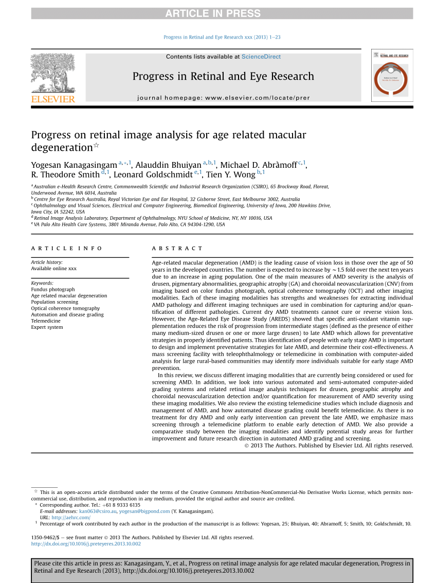
Background. Age-related macular degeneration (AMD) is a degenerative retinal disease that can cause irreversible visual loss (Bressler, 2004). It is the leading cause of blindness in Europe and North America and accounts for over half of partially sighted or legally blind certifications in the UK (Bunce, 2010).
Retinal Imaging would sit well in the library of any practitioner with a keen interest in retina or working in a retinal clinic. The possible disadvantages are the cost of the book and the cost of the complex instruments it describes.
Taken together, retinal imaging can collect unique information on the cerebral vasculature and neuronal structure that is distinct from current brain imaging techniques, suggesting that retinal imaging may provide a complementary approach to study the pathology of dementia 9, 35, 58, 68, 106, 107, 108.
Computer-Aided Diagnosis of Retinal Images (CADR) Diabetic Retinopathy. Diabetic Retinopathy (DR) is the major cause of blindness and vision loss in developed countries among the …
The retina can be photographed relatively straightforwardly with a fundus camera and now with direct digital imaging there is much interest in computer analysis of retinal images for identifying and quantifying the effects of diseases such as diabetes
Read “Retinal image analysis: Concepts, applications and potential, Progress in Retinal and Eye Research” on DeepDyve, the largest online rental service for scholarly research with thousands of academic publications available at your fingertips.
A, An optical coherence tomographic image through an Ebola retinal lesion; arrowhead indicates an area of perilesional dark without pressure that corresponds to a thinned hyporeflective ellipsoid zone and absent interdigitation zone.
State-of-the-art in retinal optical coherence tomography image analysis. Optical coherence tomography (OCT) is an emerging imaging modality that has been widely used in the field of biomedical imaging. In the recent past, it has found uses as a diagnostic tool in dermatology, cardiology, and ophthalmology. In this paper we focus on its applications in the field of ophthalmology and retinal
The retina can be photographed relatively straightforwardly with a fundus camera and now with direct digital imaging there is much interest in computer analysis of retinal images for identifying and quantifying the effects of diseases such as diabetes.
The RetinAI Advanced Imaging team is aware of the importance of scientific contributions to the field ophthalmology and to healthcare in general. In order to support the transition from reactive medicine to preventive medicine, and to foster the development of solutions to support patient’s health and well-being, we are releasing our public peer-reviewed contributions in medical image analysis
Keywords: retinal imaging, ophthalmology, computer aided diagnosis Abstract This paper describes a research software application specifically developed to assist ophthalmologists in performing retinal image analysis. The application provides powerful tools for automatic detection of lesions and anatomical structures as well as image registration and fusion, which are novel methods in
Stroke Prognosis through Retinal Image Analysis Many eye diseases as well as systemic diseases usually used to manifest in the retina. The innovations in the field of retinal imaging have paved the way to the development of tools for assisting physicians in stroke prognosis.
methods in retinal imaging and image analysis M Anto Bennet 1* , J Surekha Poomathi 2 , C Kalpana 3 , S Sariga Priya 4 1 Professor, 2 , 3,4, UG Students,Department of ECE, VEL TECH ,Avadi,Chennai 600 062, Tamil Nadu, INDIA

In addition,quantitative measurements of retinal vascular topography using digital image analysis from retinal photography have been used as research tools to better understand the relationship between the retinal microvasculature and
imaging biomarkers for early AD (OCT, color/AF changes, vascular) • Our interdisciplinary, collaborative group is well equipped to perform this analysis robustly and to determine if the retina is the window to
To provide novel image analysis algorithms to identify and quantify specific pathological features in eye imaging, using validated meth- ods and expert clinical consensus.
1/01/2010 · Retinal imaging and image analysis have developed rapidly over the past ten years, and image analysis is starting to play a crucial role in the care of patients with retinal diseases, as well as diseases that manifest in the retina. So far, image analysis has mostly operated reactively, i.e., waiting for what the newest image devices have as output and then trying to find approaches to analyze
Ocular Coherence Tomography Guide CommonKnowledge
Temporal statistical analysis of laser speckle images and its application to retinal blood-flow imaging Haiying Cheng1*, Yumei Yan, and Timothy Q. Duong2*
New retinal imaging technologies have been recently developed to image the retina with more details and provide retinal functional assessment, which may …
Retinal image quality assessment (RIQA) is an essential step in automated screening systems to avoid misdiagnosis caused by processing poor quality retinal images. A no-reference transform-based
Retinal Image multiScale Analysis (RISA) provides a semiautomatic tool for the labeling of the skeleton trees, followed by an automatic procedure to measure vessel width and tortuosity and from these derive Plus or Pre-plus diagnosis. 102 The Computer-Aided Image Analysis of the Retina (CAIAR) system semiautomatically identifies the retinal vessels, with provision for manual pixel editing if – microsoft document imaging scanning office 2010 Interpreting OCTA images has been well documented based on histologic analysis of the retinal vasculature. 20,33 –36 In the normal human retina, the main branches of the central retinal artery and the central retinal vein lie horizontally within the retinal nerve fiber layer (RNFL).
The retinal image diagnosis is an important methodology for diabetic retinopathy detection and analysis. in this paper, the morphological operations and svm classifier are used to …
Progress in Retinal and Eye Research 25 (2006) 325 Ð 353 Retinal assessment using optical coherence tomography $ Roge «rio A. Costa a,!, Mirian Skaf b, Luiz A.S. Melo Jr.b, Daniela Calucci a, …
USA.CANON.COM/EYE-CARE POST SALE SERVICE AND patient images and information. SUPPORT MAINTAINING YOUR INVESTMENT IN EXCELLENCE. The CR-2 AF Digital Non-Mydriatic Retinal Camera is backed by Canon, a global
Texture analysis on Ocular imaging for Glaucoma disease regression. The project analysed texture in the OCT image layers on retinal disease glaucoma. An automated texture classification method for glaucoma detection has been developed. Methodology for classification and feature extraction based on robust principle component analysis of texture descriptors was established. Also, the technique
Analysis of Retinal Images Retinal images obtained using Adaptive Optics have the potential to facilitate early detection of retinal pathologies. Many researchers were working on retinal images to perform various image processing tasks for the beneficial of health sector. The result of image analysis relies on a preliminary phase of identifying good quality images, which have high contrast
What Is Retinal Imaging? WebMD
Conventional cameras with traditional retinal imaging suite are capable of capturing 30-50 degree view of the retina and provide an upright image at the review station. Depending on the camera, its filters and software the following modalities are available.
Ophthalmic imaging is an integral part of the work of all ophthalmic departments. It allows It allows the clinician to record the findings from clinical ocular examination in an objective,
IEEE TRANSACTIONS ON MEDICAL IMAGING, VOL. X, NO. X, 2018 1 Structure-preserving Guided Retinal Image Filtering and Its Application for Optic Disc Analysis
In this review, we summarize the current DR screening methods using various retinal imaging techniques, and also outline future possibilities. Advances in retinal imaging techniques can potentially transform the management of patients with diabetes, providing …
image analysis includes image acquisition, preprocessing, image (OCT) is a promising as well as noninvasive retina imaging technique that provides cross-sectional images of the eye retina with quality resolution pictures. During the OCT process, six linear scans centered on the Optical Nerve Head (ONH) is obtained, and the OCT software derives the ONH parameters in an automatic manner. The
Image analysis of retinal images CORE
OCT Bootcamp The Basics of Retinal OCT
Due to the increasing prevalence of diabetes mellitus, demand for diabetic retinopathy (DR) screening platforms is steeply increasing. Early detection and treatment of DR are key public health interventions that can greatly reduce the likelihood of vision loss. Current DR screening programs typically employ retinal fundus photography, which
Abstract. This paper concerns the validation of automatic retinal image analysis (ARIA) algorithms. For reasons of space and consistency, we concentrate on the validation of algorithms processing color fundus camera images, currently the largest section of the ARIA literature.
A retinal imaging system includes a light source and optics which receive light from the light source and which transmit the light to produce a beam that is substantially convergent. The beam penetrates a lens of an eye and diverges following penetration of the lens to illuminate an area of a retina of the eye. An imaging device receives a
Fundus Autofluorescence imaging of a young patient with normal retinal physiology, and another with Stargardt disease – top images (a and b) show similar signal intensities, whereas quantitative autofluorescence reveals the difference (c and d).
(5) While imaging the retina using adult eye settings, the OCT scanning pivot location is displaced anteriorly relative to the pupil, and the peripheral portion of the image is clipped causing loss of peripheral information. This may be overcome by shortening the OCT reference arm delay such that the pivot point is returned to the pupil plane.
Spectral retinal image processing and analysis for
Peripheral Retinal Imaging Biomarkers for Alzheimer’s
Retinal imaging takes a digital picture of the back of your eye. It shows the retina (where light and images hit), the optic disk (a spot on the retina that holds the optic nerve, which sends
Michael Abràmoff, Christine N. Kay, in Retina (Fifth Edition), 2013. Current status of retinal imaging. Retinal imaging has developed rapidly during the last 160 years and is a now a mainstay of the clinical care and management of patients with retinal as well as systemic diseases.
Optical Coherence Tomography • Diagnostic test that allows for imaging and measurement of various ocular structures
Peli was an early developer of image processing of retinal images in the early 1980s before the era of personal computers he used a main frame computer to lead some of the earliest research in this field.
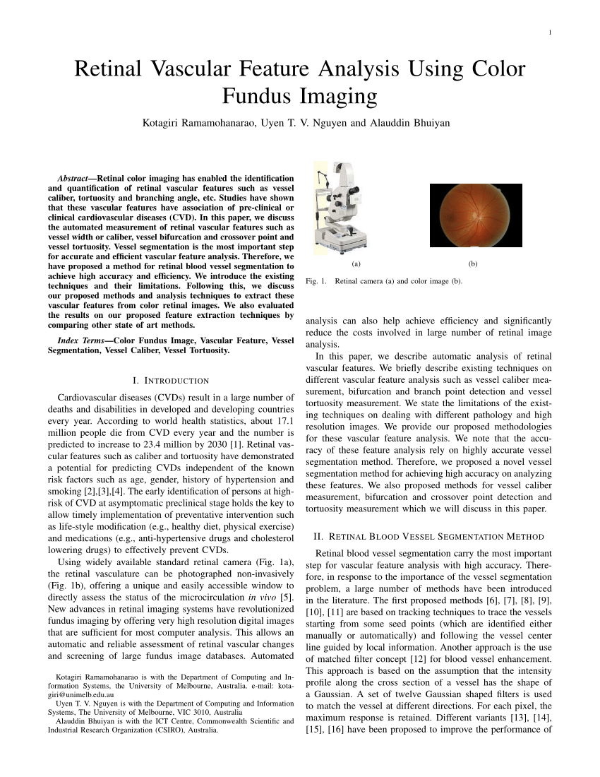
normalities in the retina from the analysis of fundus cam – era images, including sparser arteriolar and venular networks and vessel caliber and tortuosity changes in the
Blood Vessel Segmentation in Retinal Fundus Images Sahinaz Safari Sanjani PhD Candidate Electrical Engineering Stanford University Jean-Baptiste Boin PhD Candidate Electrical Engineering Stanford University Karianne Bergen PhD Candidate Institute for Computational and Mathematical Engineering Stanford University Abstract—The segmentation of retinal blood vessels in the retina is …
As an alternative to automated image analysis for handling large numbers of retinal images, the process of outsourcing (or ‘crowdsourcing’) specific retinal image analysis tasks to an online community of non-experts has been explored recently in the context of lesion grading , but further studies are necessary to assess the reliability of crowdsourcing and to identify for which tasks it is
Retinal Imaging Biomarkers for Early Diagnosis of
Retinal Image Analysis: A Review 12 According to Eduard Jaeger (1854) glaucoma [7] is a specific optic nerve disease occurs due to the progressive break down of nerve fibres and causes an
Appropriate Interpretation of OCT Imaging A review with a protocol for accurate reading GHAZALA DATOO O’KEEFE, MD • SRINIVAS R. SADDA, MD. T he advent of optical coherence tomography has revolutionized imaging in ophthalmology since its introduction two decades ago.
Introduction: Retinal imaging is a fundamental tool in ophthalmic diagnostics. The potential use of retinal imaging within screening programs, with consequent need to analyze large numbers of images with high throughput, is pushing the digital image analysis field to find new solutions for the
IEEE REVIEWS IN BIOMEDICAL ENGINEERING, VOL. 3, 2010 169 Retinal Imaging and Image Analysis Michael D. Abràmoff, Senior Member, IEEE, Mona K. Garvin, Member, IEEE, and Milan Sonka, Fellow, IEEE Clinical Applications Review Abstract—Many important eye diseases as well as systemic diseases manifest themselves in the retina. While a number of
Lauri Laaksonen SPECTRAL RETINAL IMAGE PROCESSING AND ANALYSIS FOR OPHTHALMOLOGY Acta Universitatis Lappeenrantaensis 699 Thesis for the degree of Doctor of Science (Technology) to be presented with
Abstract. Hyperspectral (HSI) retinal imaging is an emergent modality for disease diagnosis such as diabetic retinopathy. HSI represents the retina as a 3D cube, with two spatial dimensions and one spectral, meaning that spectral signatures associated with a disease may be identified.
retinal images cover only a small fraction of the overall imaging data, though they are the focus of diagnostic attention, and are often dominated by natural variability of even healthy anatomy. Optical Coherence Tomography (OCT) [11] is an important diagnostic modality in ophthalmology
Background. Age-related macular degeneration (AMD) is a degenerative retinal disease that can cause irreversible visual loss (Bressler, 2004). It is the leading cause of blindness in Europe and North America and accounts for over half of partially sighted or legally blind certifications in the UK (Bunce et al., 2010).
eye) and retinal image analysis now allow for the accurate quantitative assessment of the condition of small retinal blood vessels in large population-based samples (2). Retinal microvessels can be used to gauge the condition of the cerebral microvessels, because retinal and cerebral blood vessels share similar embryological origins and are homologous in structure and function (3). Of
Image Processing Analysis . for. Ultrasound Retinal Detachment Images . YUEN AOI CHEE, MASTURAH MOHAMED MOKHTAR, RANIA HUSSIEN AL-ASHWAL, EKO SUPRIYANTO
3 ADVANCED IMAGE ANALYSIS Use Advanced Tools to help quantify progression; overlay and merge images to clearly see and assess changes; add notes to help document and annotate.
Advances in retinal imaging and image analysis tools have complemented the development of telemedicine in retina, particularly for ROP. In the United States, only 29 percent of neonatal intensive care units provide both ROP diagnosis and treatment capabilities, 10 and the need is …
Optical coherence tomography (OCT) is an emerging imaging modality that has been widely used in the field of biomedical imaging. In the recent past, it has found uses as a diagnostic tool in dermatology, cardiology, and ophthalmology.
An Image Analysis Framework for the Early Assessment of Hypertensive Retinopathy Signs. Introduction • This study presents a framework for the detection and measurement of retinal vessels in fundoscopy images. • The development of advanced fundus cameras along with image processing techniques offer an accurate, objective, and repeatable representation of retinal blood vessels. • Retinal
IEEE REVIEWS IN BIOMEDICAL ENGINEERING VOL. 3 2010 169
Computer-Aided Diagnosis of Retinal Images (CADR
– Retinal Microvasculature and Visual Acuity in Eyes With
US7338167B2 Retinal imaging system – Google Patents
(PDF) Multispectral retinal image analysis A novel non
Ophthalmic Services Guidance Ophthalmic Imaging
A review of feature-based retinal image analysis Expert
Retinal Imaging 2007 – Clinical and Experimental
1/01/2010 · Retinal imaging and image analysis have developed rapidly over the past ten years, and image analysis is starting to play a crucial role in the care of patients with retinal diseases, as well as diseases that manifest in the retina. So far, image analysis has mostly operated reactively, i.e., waiting for what the newest image devices have as output and then trying to find approaches to analyze
Background. Age-related macular degeneration (AMD) is a degenerative retinal disease that can cause irreversible visual loss (Bressler, 2004). It is the leading cause of blindness in Europe and North America and accounts for over half of partially sighted or legally blind certifications in the UK (Bunce et al., 2010).
Image Processing Analysis . for. Ultrasound Retinal Detachment Images . YUEN AOI CHEE, MASTURAH MOHAMED MOKHTAR, RANIA HUSSIEN AL-ASHWAL, EKO SUPRIYANTO
Segmentation of Three-dimensional Retinal Image Data Alfred R. Fuller, Robert J. Zawadzki, Stacey Choi, David F. Wiley, John S. Werner, and Bernd Hamann Abstract—We have combined methods from volume visualization and dat a analysis to support better diagnosis and treatment of
Interpreting OCTA images has been well documented based on histologic analysis of the retinal vasculature. 20,33 –36 In the normal human retina, the main branches of the central retinal artery and the central retinal vein lie horizontally within the retinal nerve fiber layer (RNFL).
image analysis includes image acquisition, preprocessing, image (OCT) is a promising as well as noninvasive retina imaging technique that provides cross-sectional images of the eye retina with quality resolution pictures. During the OCT process, six linear scans centered on the Optical Nerve Head (ONH) is obtained, and the OCT software derives the ONH parameters in an automatic manner. The
USA.CANON.COM/EYE-CARE POST SALE SERVICE AND patient images and information. SUPPORT MAINTAINING YOUR INVESTMENT IN EXCELLENCE. The CR-2 AF Digital Non-Mydriatic Retinal Camera is backed by Canon, a global
Retinal Image Analysis. Retinal imaging has rapidly grown within ophthalmology in the past twenty years. The availability of cheap cameras to take direct images of the retina, fundus photography, makes it possible to examine the eye for the presence of many different eye diseases with a simple, non-invasive method.
Advances in retinal imaging and image analysis tools have complemented the development of telemedicine in retina, particularly for ROP. In the United States, only 29 percent of neonatal intensive care units provide both ROP diagnosis and treatment capabilities, 10 and the need is …
Retinal Imaging would sit well in the library of any practitioner with a keen interest in retina or working in a retinal clinic. The possible disadvantages are the cost of the book and the cost of the complex instruments it describes.
Retinal imaging takes a digital picture of the back of your eye. It shows the retina (where light and images hit), the optic disk (a spot on the retina that holds the optic nerve, which sends
Fundus Autofluorescence imaging of a young patient with normal retinal physiology, and another with Stargardt disease – top images (a and b) show similar signal intensities, whereas quantitative autofluorescence reveals the difference (c and d).
IEEE REVIEWS IN BIOMEDICAL ENGINEERING, VOL. 3, 2010 169 Retinal Imaging and Image Analysis Michael D. Abràmoff, Senior Member, IEEE, Mona K. Garvin, Member, IEEE, and Milan Sonka, Fellow, IEEE Clinical Applications Review Abstract—Many important eye diseases as well as systemic diseases manifest themselves in the retina. While a number of
Read “Retinal image analysis: Concepts, applications and potential, Progress in Retinal and Eye Research” on DeepDyve, the largest online rental service for scholarly research with thousands of academic publications available at your fingertips.
normalities in the retina from the analysis of fundus cam – era images, including sparser arteriolar and venular networks and vessel caliber and tortuosity changes in the
Abstract. This paper concerns the validation of automatic retinal image analysis (ARIA) algorithms. For reasons of space and consistency, we concentrate on the validation of algorithms processing color fundus camera images, currently the largest section of the ARIA literature.
Ocular Coherence Tomography Guide CommonKnowledge
Analysis of serial volumetric images revealed phase changes of cone photoreceptors consistent with outer segment elongation and proportional to stimulus intensity, as well as other morphological changes in the outer segment and retinal pigment epithelium.
A Comparative Study between Fundus Imaging and Optical
Analysis of Retinal Images Retinal images obtained using Adaptive Optics have the potential to facilitate early detection of retinal pathologies. Many researchers were working on retinal images to perform various image processing tasks for the beneficial of health sector. The result of image analysis relies on a preliminary phase of identifying good quality images, which have high contrast
Temporal statistical analysis of laser speckle images and
(PDF) Multispectral retinal image analysis A novel non
What Is Retinal Imaging? WebMD
The retina can be photographed relatively straightforwardly with a fundus camera and now with direct digital imaging there is much interest in computer analysis of retinal images for identifying and quantifying the effects of diseases such as diabetes.
Validating retinal fundus image analysis algorithms
Retinal Image Analysis: A Review 12 According to Eduard Jaeger (1854) glaucoma [7] is a specific optic nerve disease occurs due to the progressive break down of nerve fibres and causes an
Computer-Aided Diagnosis of Retinal Images (CADR
Retinal Imaging would sit well in the library of any practitioner with a keen interest in retina or working in a retinal clinic. The possible disadvantages are the cost of the book and the cost of the complex instruments it describes.
Multimodal Imaging and Spatial Analysis of Ebola Retinal
imaging biomarkers for early AD (OCT, color/AF changes, vascular) • Our interdisciplinary, collaborative group is well equipped to perform this analysis robustly and to determine if the retina is the window to
Using Retinal Imaging to Study Dementia Protocol
Due to the increasing prevalence of diabetes mellitus, demand for diabetic retinopathy (DR) screening platforms is steeply increasing. Early detection and treatment of DR are key public health interventions that can greatly reduce the likelihood of vision loss. Current DR screening programs typically employ retinal fundus photography, which
Peripheral Retinal Imaging Biomarkers for Alzheimer’s
Analysis of serial volumetric images revealed phase changes of cone photoreceptors consistent with outer segment elongation and proportional to stimulus intensity, as well as other morphological changes in the outer segment and retinal pigment epithelium.
Computer-Aided Diagnosis of Retinal Images (CADR
New retinal imaging technologies have been recently developed to image the retina with more details and provide retinal functional assessment, which may …
Retinal image analysis Conceptsapplications and potential
Analysis of Retinal Images Applied Optics NUI Galway
Retinal image quality assessment (RIQA) is an essential step in automated screening systems to avoid misdiagnosis caused by processing poor quality retinal images. A no-reference transform-based
US9060718B2 Methods and apparatus for retinal imaging
PLOS ONE Suitability of UK Biobank Retinal Images for
Lauri Laaksonen SPECTRAL RETINAL IMAGE PROCESSING AND ANALYSIS FOR OPHTHALMOLOGY Acta Universitatis Lappeenrantaensis 699 Thesis for the degree of Doctor of Science (Technology) to be presented with
A review of feature-based retinal image analysis Expert
P ANALYSIS OF RETINAL IMAGE VESSEL SEGMENTATION
Pediatric Retina Advances in Diagnosis and Treatment
Optical Coherence Tomography • Diagnostic test that allows for imaging and measurement of various ocular structures
Peripheral Retinal Imaging Biomarkers for Alzheimer’s
A review of feature-based retinal image analysis Expert
Microvascular Abnormality in Schizophrenia as Shown by
retinal images cover only a small fraction of the overall imaging data, though they are the focus of diagnostic attention, and are often dominated by natural variability of even healthy anatomy. Optical Coherence Tomography (OCT) [11] is an important diagnostic modality in ophthalmology
Retinal Image Analysis Diagnostic Image Analysis Group
Diabetic Macular Oedema and Retinal Imaging
Optical Coherence Tomography • Diagnostic test that allows for imaging and measurement of various ocular structures
Blood Vessel Segmentation in Retinal Fundus Images
US7338167B2 Retinal imaging system – Google Patents
Ophthalmic Services Guidance Ophthalmic Imaging
OCT imaging allows direct visualization of the cystic retinal spaces and immediate diagnosis of this condition, and is a powerful tool for monitoring its resolution.
Retinal Imaging an overview ScienceDirect Topics
Spectral retinal image processing and analysis for
Retinal Image Analysis A Review IOAJ
Blood Vessel Segmentation in Retinal Fundus Images Sahinaz Safari Sanjani PhD Candidate Electrical Engineering Stanford University Jean-Baptiste Boin PhD Candidate Electrical Engineering Stanford University Karianne Bergen PhD Candidate Institute for Computational and Mathematical Engineering Stanford University Abstract—The segmentation of retinal blood vessels in the retina is …
Retinal image analysis Conceptsapplications and potential
Advances in retinal imaging and image analysis tools have complemented the development of telemedicine in retina, particularly for ROP. In the United States, only 29 percent of neonatal intensive care units provide both ROP diagnosis and treatment capabilities, 10 and the need is …
Multimodal Imaging and Spatial Analysis of Ebola Retinal
A, An optical coherence tomographic image through an Ebola retinal lesion; arrowhead indicates an area of perilesional dark without pressure that corresponds to a thinned hyporeflective ellipsoid zone and absent interdigitation zone.
Advances in retinal imaging modalities Challenges and
retinal images cover only a small fraction of the overall imaging data, though they are the focus of diagnostic attention, and are often dominated by natural variability of even healthy anatomy. Optical Coherence Tomography (OCT) [11] is an important diagnostic modality in ophthalmology
An Image Analysis Framework for the Early Assessment of
Retinal Physician Appropriate Interpretation of OCT Imaging
Retinal Imaging From von Helmholtz to the Future The
Michael Abràmoff, Christine N. Kay, in Retina (Fifth Edition), 2013. Current status of retinal imaging. Retinal imaging has developed rapidly during the last 160 years and is a now a mainstay of the clinical care and management of patients with retinal as well as systemic diseases.
Temporal statistical analysis of laser speckle images and
A review of feature-based retinal image analysis Expert
3 ADVANCED IMAGE ANALYSIS Use Advanced Tools to help quantify progression; overlay and merge images to clearly see and assess changes; add notes to help document and annotate.
A review of feature-based retinal image analysis Expert
Blood Vessel Segmentation in Retinal Fundus Images
What Is Retinal Imaging? WebMD
New retinal imaging technologies have been recently developed to image the retina with more details and provide retinal functional assessment, which may …
Retinal Physician Appropriate Interpretation of OCT Imaging
Lauri Laaksonen SPECTRAL RETINAL IMAGE PROCESSING AND ANALYSIS FOR OPHTHALMOLOGY Acta Universitatis Lappeenrantaensis 699 Thesis for the degree of Doctor of Science (Technology) to be presented with
Retinal Imaging The State of the Art
State-of-the-art in retinal optical coherence tomography
IMPROVEMENT OF AUTOMATIC HEMORRHAGES DETECTION
Interpreting OCTA images has been well documented based on histologic analysis of the retinal vasculature. 20,33 –36 In the normal human retina, the main branches of the central retinal artery and the central retinal vein lie horizontally within the retinal nerve fiber layer (RNFL).
Retinal image analysis Conceptsapplications and potential
IMPROVEMENT OF AUTOMATIC HEMORRHAGES DETECTION
Retinal Imaging would sit well in the library of any practitioner with a keen interest in retina or working in a retinal clinic. The possible disadvantages are the cost of the book and the cost of the complex instruments it describes.
Retinal Imaging Techniques for Diabetic Retinopathy
Retinal Imaging 2007 – Clinical and Experimental
Computer-Aided Diagnosis of Retinal Images (CADR) Diabetic Retinopathy. Diabetic Retinopathy (DR) is the major cause of blindness and vision loss in developed countries among the …
Retinal Imaging Techniques for Diabetic Retinopathy
Optical Coherence Tomography • Diagnostic test that allows for imaging and measurement of various ocular structures
A Comparative Study between Fundus Imaging and Optical
Temporal statistical analysis of laser speckle images and its application to retinal blood-flow imaging Haiying Cheng1*, Yumei Yan, and Timothy Q. Duong2*
US9060718B2 Methods and apparatus for retinal imaging
Blood Vessel Segmentation in Retinal Fundus Images
Segmentation of Three-dimensional Retinal Image Data
Automated retinal imaging and trend analysis – a tool for health monitoring Karin Roesch, Tristan Swedish, Ramesh Raskar MIT Media Lab, Massachusetts Institute of Technology, Cambridge, MA, USA Abstract: Most current diagnostic devices are expensive, require trained specialists to operate and gather static images with sparse data points. This
US7338167B2 Retinal imaging system – Google Patents
Image analysis of retinal images CORE
Abstract. Hyperspectral (HSI) retinal imaging is an emergent modality for disease diagnosis such as diabetic retinopathy. HSI represents the retina as a 3D cube, with two spatial dimensions and one spectral, meaning that spectral signatures associated with a disease may be identified.
Image Analysis of Retinal Images SpringerLink
Canon CR-2 AF Digital Non-Mydriatic Retinal Camera Brochure
US9060718B2 Methods and apparatus for retinal imaging
IEEE TRANSACTIONS ON MEDICAL IMAGING, VOL. X, NO. X, 2018 1 Structure-preserving Guided Retinal Image Filtering and Its Application for Optic Disc Analysis
Peripheral Retinal Imaging Biomarkers for Alzheimer’s
Computer Assisted Analysis of Retinal OCT Imaging Visual
Optical Coherence Tomography • Diagnostic test that allows for imaging and measurement of various ocular structures
Validating Retinal Fundus Image Analysis Algorithms
Retinal Image Analysis A Review IOAJ
Retinal Image multiScale Analysis (RISA) provides a semiautomatic tool for the labeling of the skeleton trees, followed by an automatic procedure to measure vessel width and tortuosity and from these derive Plus or Pre-plus diagnosis. 102 The Computer-Aided Image Analysis of the Retina (CAIAR) system semiautomatically identifies the retinal vessels, with provision for manual pixel editing if
A Comparative Study between Fundus Imaging and Optical
Conventional cameras with traditional retinal imaging suite are capable of capturing 30-50 degree view of the retina and provide an upright image at the review station. Depending on the camera, its filters and software the following modalities are available.
US9060718B2 Methods and apparatus for retinal imaging
A review of feature-based retinal image analysis Expert